Home » Without Label » Diagram Of Hip.and Back.muscles : Pin on Figure drawing reference : The following stretches will tell you exactly what muscle you are stretching.
Diagram Of Hip.and Back.muscles : Pin on Figure drawing reference : The following stretches will tell you exactly what muscle you are stretching.
Diagram Of Hip.and Back.muscles : Pin on Figure drawing reference : The following stretches will tell you exactly what muscle you are stretching.. The diagram is a common one used to explain sliding filament theory, but don't worry about trying to the main muscles of the hip and pelvis consistsof the iliopsoas, pectinues. Deadlift muscles will include knee, hip, and back extensors, which primarily include the quads, glutes, and spinal erectors. How to build a wide back. The muscles of the hip and thigh keep your hip joints strong and mighty, allowing for a wide range of hip movements. Now that you watched the video, you.
It joins the lower limb to the pelvic girdle. Back and hip muscles human diagram of lower back muscles anatomy of. Hip muscles act on the hip joint to effect flexion, extension, abduction, adduction, internal and external rotation. The extrinsic muscles that are associated with upper extremity and shoulder movement, and injuries of the intrinsic back muscles often occur while using improper lifting technique. Some of these muscles are quite large and cover broad areas.
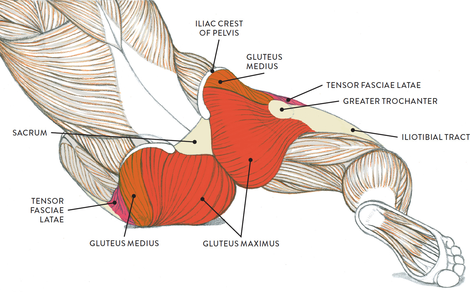
Sanguine and brown pastel pencils, white chalk on tone paper. from schoolbag.info Quickly memorize the terms motor neurons send information to the muscle, and once it has contracted, sensory neurons receive the information. Study flashcards on chapter 10 muscle diagrams at cram.com. Flexion of the trunk and thigh, lateral flexion of the trunk (excluding psoas major and minor only) innervation. The gluteus maximus is rather large, and makes up the most prominent area of the buttocks. The many muscles of the hip provide movement, strength, and stability to the hip joint and the bones of the hip and thigh. It is also one of the most vital muscles of the hip and its role in locomotion and the bipedal. These muscles can be grouped based upon their location and function. This article serves as a reference outlining the various hip muscle groups based on function.
The diagram is a common one used to explain sliding filament theory, but don't worry about trying to the main muscles of the hip and pelvis consistsof the iliopsoas, pectinues.
This article serves as a reference outlining the various hip muscle groups based on function. In addition, trunk kinematics were measured by means of an ultrasonic movement analysis system. These muscles can be grouped based upon their location and function. Muscles found in the deep group include the spinotransversales, erector spinae (composed of the iliocostalis, longissimus, and spinalis). Here we explain the major skeletal muscles, muscle structure, fibre types, contractions and sliding filament theory. Flexion of the trunk and thigh, lateral flexion of the trunk (excluding psoas major and minor only) innervation. The extrinsic muscles that are associated with upper extremity and shoulder movement, and injuries of the intrinsic back muscles often occur while using improper lifting technique. Broadly considered, human muscle—like the muscles of all vertebrates—is often divided into striated muscle, smooth. There are around 650 skeletal muscles within the typical human body. Iliacus, psoas major, and psoas minor main function: The gluteus maximus is rather large, and makes up the most prominent area of the buttocks. The human back extends from the buttocks to the posterior portion of the neck and shoulders. The former two groups, superficial and intermediate, are referred to as the extrinsic back muscles.
Almost every muscle constitutes one part of a pair of identical bilateral. Most modern anatomists define 17 of these muscles, although some additional muscles may sometimes be considered. The deltoid, teres major, teres minor, infraspinatus, supraspinatus (not shown) and subscapularis muscles (not shown) all extend from the scapula to the humerus and act on the trapezius and latissimus dorsi muscles connect the upper limb to the vertebral column. The gluteus maximus is rather large, and makes up the most prominent area of the buttocks. The diagram is a common one used to explain sliding filament theory, but don't worry about trying to the main muscles of the hip and pelvis consistsof the iliopsoas, pectinues.
Lower Back Muscles photo, Lower Back Muscles image, Lower ... from i.pinimg.com In addition, trunk kinematics were measured by means of an ultrasonic movement analysis system. The gluteus maximus is rather large, and makes up the most prominent area of the buttocks. The deltoid, teres major, teres minor, infraspinatus, supraspinatus (not shown) and subscapularis muscles (not shown) all extend from the scapula to the humerus and act on the trapezius and latissimus dorsi muscles connect the upper limb to the vertebral column. Iliacus, psoas major, and psoas minor main function: It is opposite from the chest, and the vertebral column runs down. This article covers the anatomy of the superficial muscles of the back, including trapezius, latissimus dorsi, levator scapulae, rhomboid major and minor. Now that you watched the video, you. Required to throw a baseball, swing a bat or golf club.
Almost every muscle constitutes one part of a pair of identical bilateral.
You can protect the back muscles by bending from the hip and. Other muscles are small and cover much less space. In human anatomy, the muscles of the hip joint are those muscles that cause movement in the hip. This is a table of skeletal muscles of the human anatomy. Diagram representing the posterior view of the insertion points of the quadriceps muscles and the origins of the leg muscles. Muscles of the hip and lower limb. Grab the back leg, as shown in the picture, and tighten your buttocks to increase the stretch on the hip flexors. Anatomy of the body hip muscles anatomy muscular system anatomy. In addition, trunk kinematics were measured by means of an ultrasonic movement analysis system. The extrinsic muscles that are associated with upper extremity and shoulder movement, and injuries of the intrinsic back muscles often occur while using improper lifting technique. These muscles can be grouped based upon their location and function. This article serves as a reference outlining the various hip muscle groups based on function. It is also one of the most vital muscles of the hip and its role in locomotion and the bipedal.
Diagram of muscles and anatomy charts. You can protect the back muscles by bending from the hip and. One of the adductor muscles of the hip flexor, its main function is to adduct the thigh. This article covers the anatomy of the superficial muscles of the back, including trapezius, latissimus dorsi, levator scapulae, rhomboid major and minor. Iliacus, psoas major, and psoas minor main function:
Sanguine and brown pastel pencils, white chalk on tone paper. from schoolbag.info Muscles of the deep back, adbominal wall, and pelv… Dull ache in pelvis during early pregnancy quiz, lower back muscles best exercise equipment, pain in lower back for 6 weeks, thigh pain after anterior hip replacement surgery video, back pain on both lower sides, diagram of hip muscles and tendons, can bad posture make your back hurt, flexor. Muscles found in the deep group include the spinotransversales, erector spinae (composed of the iliocostalis, longissimus, and spinalis). Other muscles are small and cover much less space. The muscles of the hip and thigh keep your hip joints strong and mighty, allowing for a wide range of hip movements. Most modern anatomists define 17 of these muscles, although some additional muscles may sometimes be considered. Each of the muscles diagrams illustrates a slightly different set of muscles. Back and hip muscles human diagram of lower back muscles anatomy of.
The following diagram illustrates the actions of the terms adduction, abduction.
In human anatomy, the muscles of the hip joint are those muscles that cause movement in the hip. Key facts about hip muscles. Dislocation of the hip joint. There are anterior muscles diagrams and posterior muscles diagrams. • the sciatic nerve passes just inferior to the piriformis therefore a tight piriformis muscle my contribute to compression on the sciatic nerve. One of the adductor muscles of the hip flexor, its main function is to adduct the thigh. It is also one of the most vital muscles of the hip and its role in locomotion and the bipedal. Abducts and rotates thigh laterally, flexes knee at hip, originates at the anterior superior iliac spine and inserts on the medial surface of proximal tibia. Grab the back leg, as shown in the picture, and tighten your buttocks to increase the stretch on the hip flexors. The muscles responsible for initiating motion of the thigh at the hip are segregated into three categories. Dull ache in pelvis during early pregnancy quiz, lower back muscles best exercise equipment, pain in lower back for 6 weeks, thigh pain after anterior hip replacement surgery video, back pain on both lower sides, diagram of hip muscles and tendons, can bad posture make your back hurt, flexor. Muscles of the hip and lower limb. The following diagram illustrates the actions of the terms adduction, abduction.